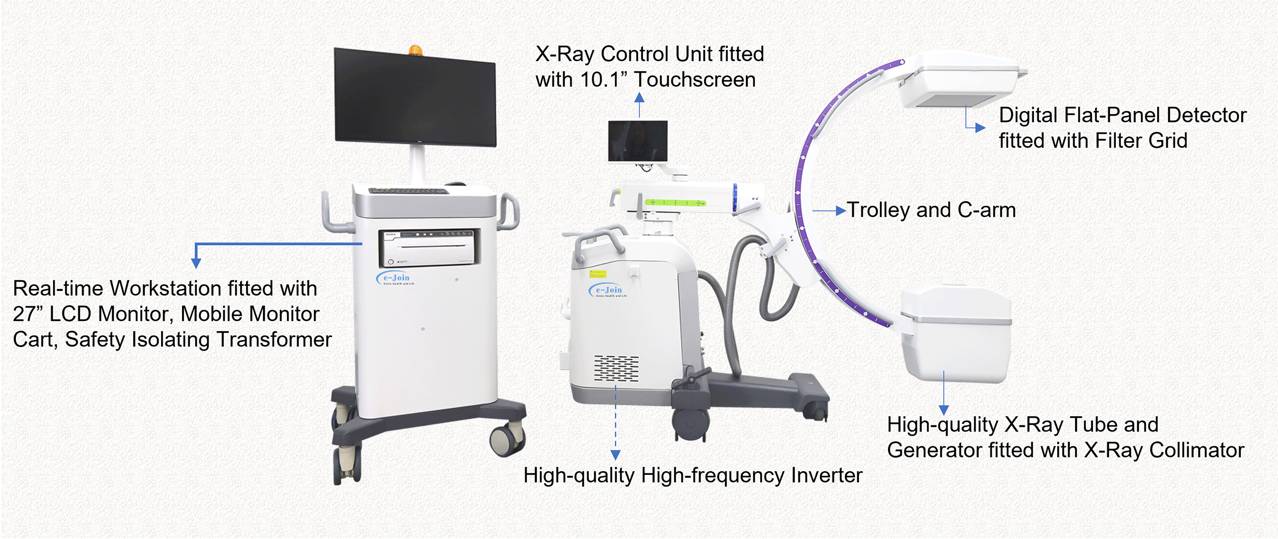
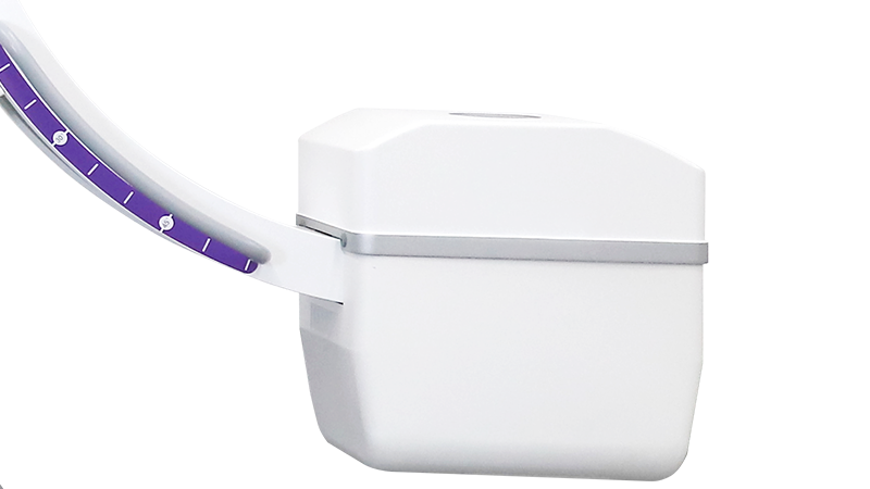
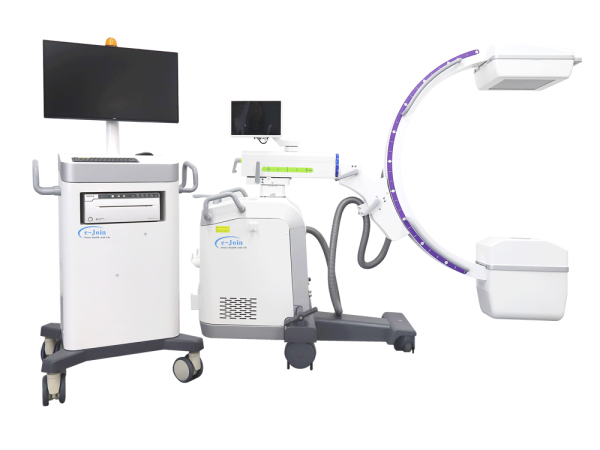
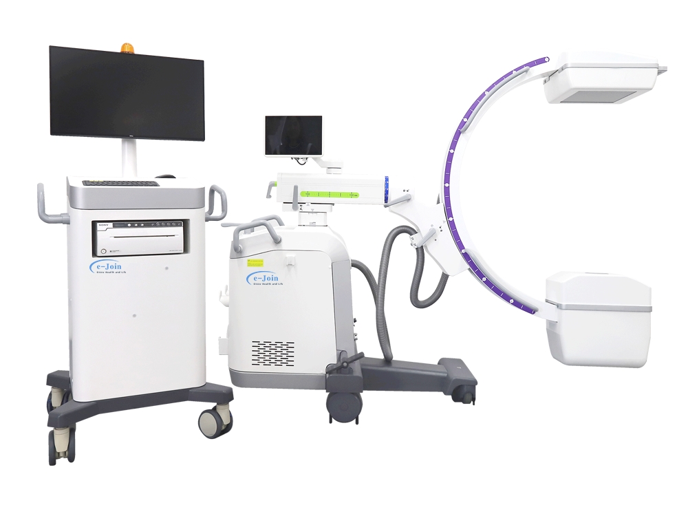
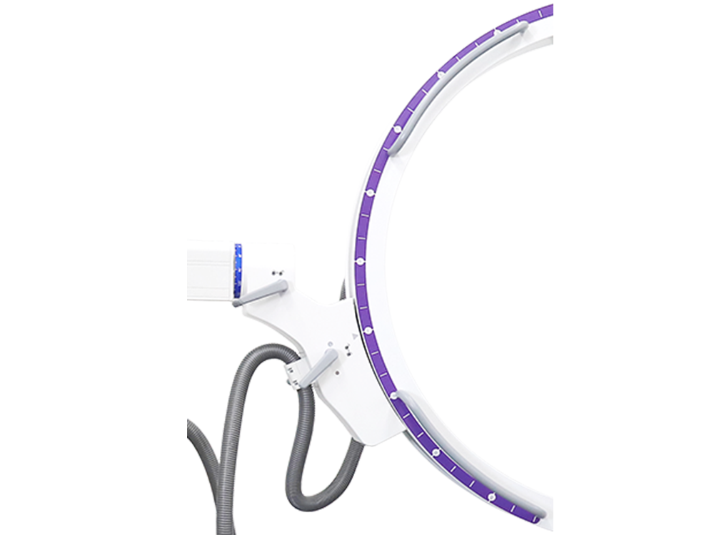
Color-coded axis and brake make operation more accurate.
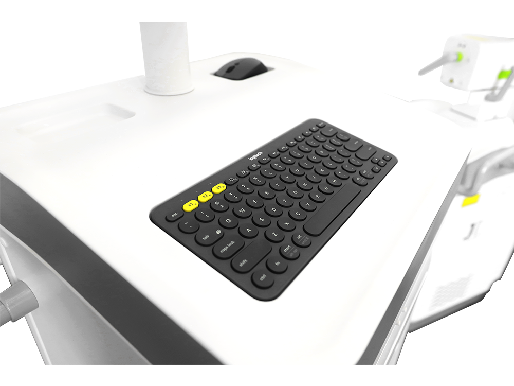
Concise control panel realizes direct operation.
Excellent C-arm self-balance design in accordance with ergonomics.
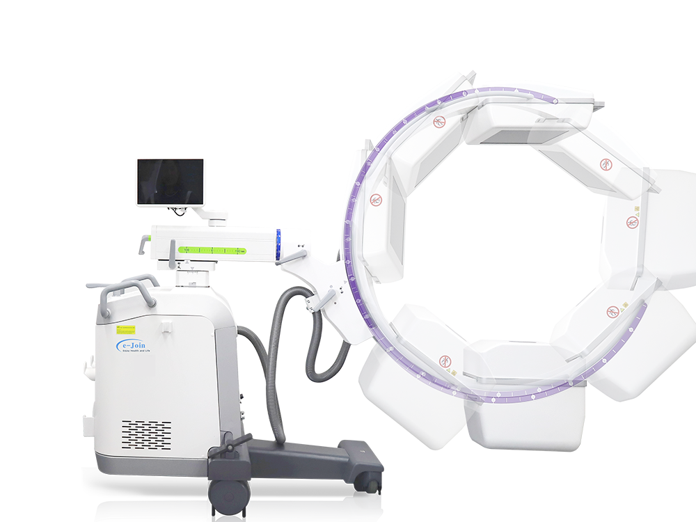
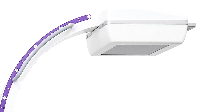
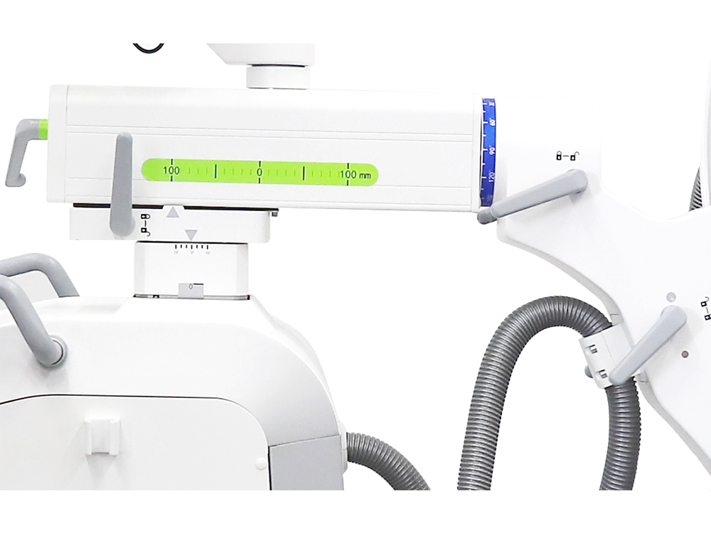
Orbital Motion
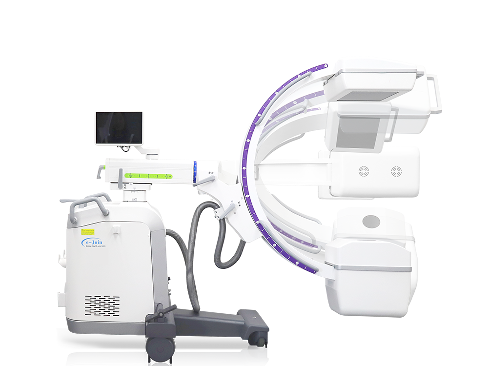
Axial Motion
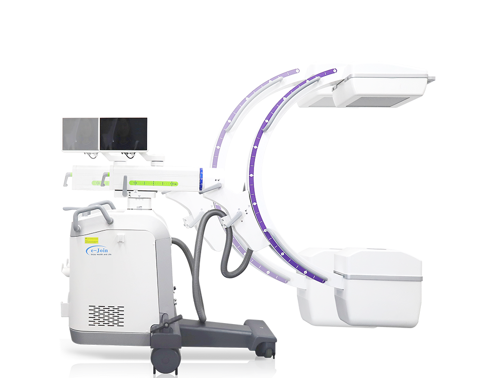
Horizontal Motion
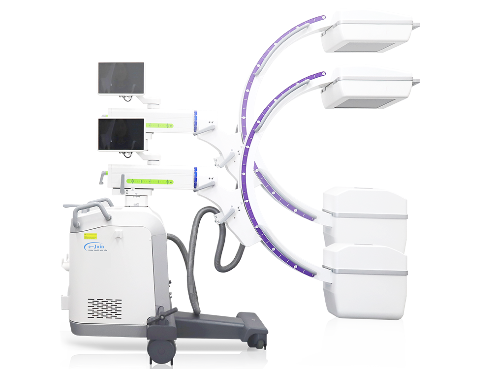
Vertical Motion
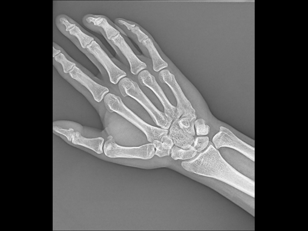
Hand
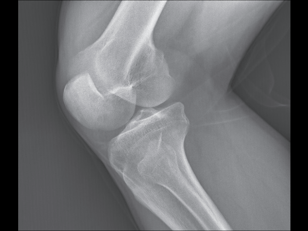
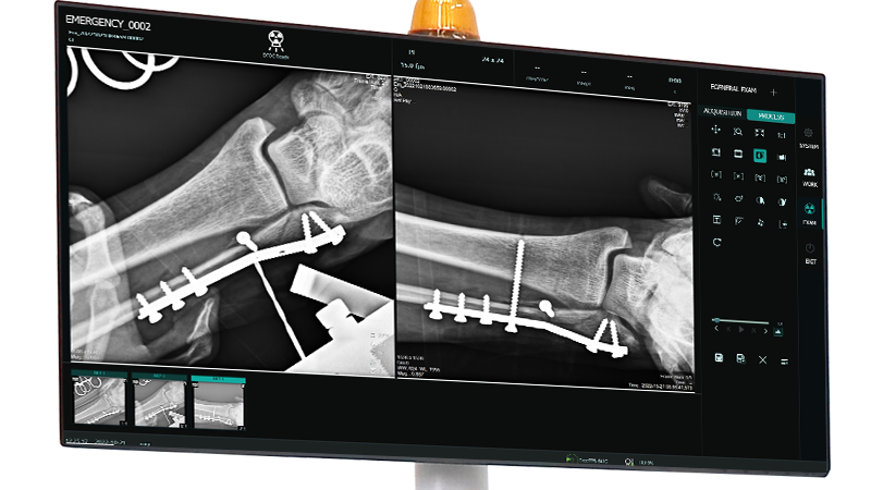
Kneecap
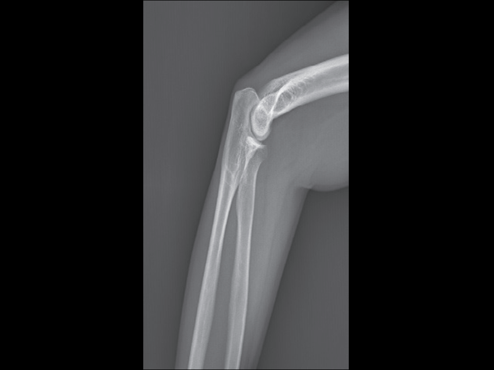
Elbow
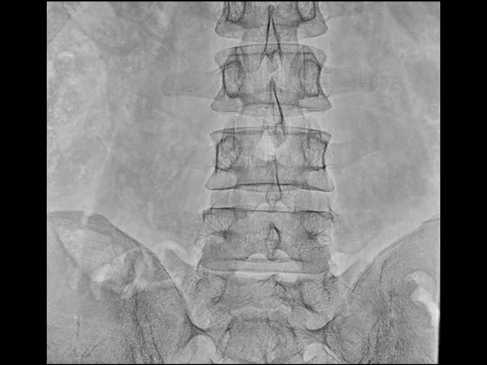
Frontal Position of Lumbar Vertebra
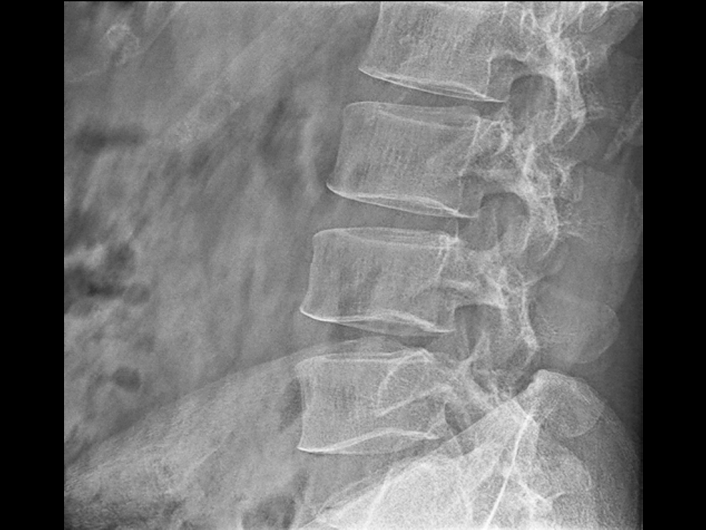
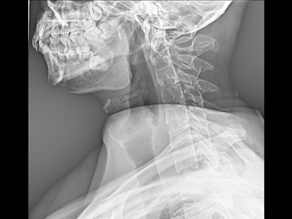
Lateral Position of Lumbar Vertebra
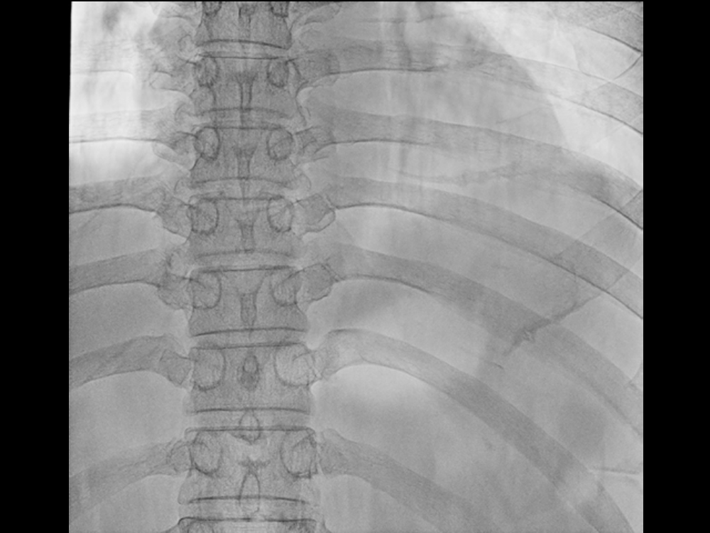
Frontal Position of Thoracic Vertebra